Home

tutta la vita diapositiva mercenario metal pdb spina folle comuni

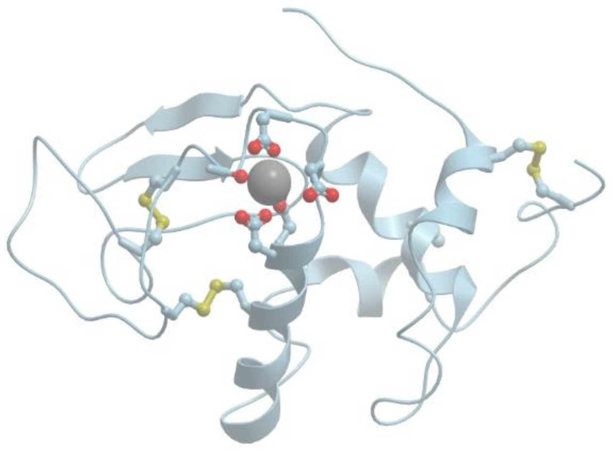
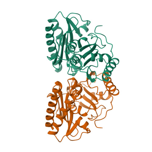
RCSB PDB - 1ELS: CATALYTIC METAL ION BINDING IN ENOLASE: THE CRYSTAL  STRUCTURE OF ENOLASE-MN2+-PHOSPHONOACETOHYDROXAMATE COMPLEX AT 2.4  ANGSTROMS RESOLUTION
RCSB PDB - 1ELS: CATALYTIC METAL ION BINDING IN ENOLASE: THE CRYSTAL STRUCTURE OF ENOLASE-MN2+-PHOSPHONOACETOHYDROXAMATE COMPLEX AT 2.4 ANGSTROMS RESOLUTION

RCSB PDB - 7KF6: Cryo-electron microscopy structure of the heavy metal  efflux pump CusA in a homogeneous binding copper(1) state
RCSB PDB - 7KF6: Cryo-electron microscopy structure of the heavy metal efflux pump CusA in a homogeneous binding copper(1) state
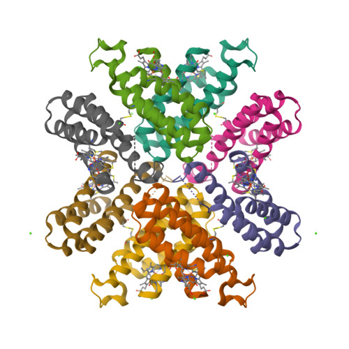
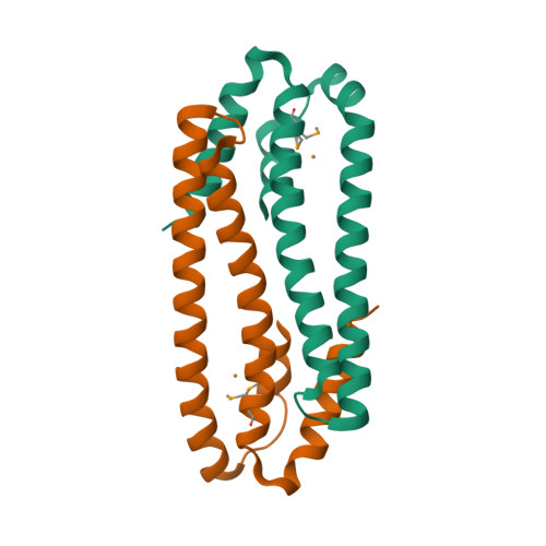
RCSB PDB - 6DHZ: Crystallographic octamer of a metal-free RIDC1 variant  bearing two disulfide bonded cysteines
RCSB PDB - 6DHZ: Crystallographic octamer of a metal-free RIDC1 variant bearing two disulfide bonded cysteines
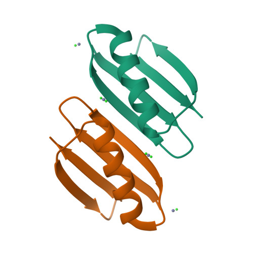
RCSB PDB - 6NL8: Crystal structure of de novo designed metal-controlled  dimer of mutant B1 immunoglobulin-binding domain of Streptococcal Protein G  (L12H, T16L, V29H, Y33H, N37L)-zinc
RCSB PDB - 6NL8: Crystal structure of de novo designed metal-controlled dimer of mutant B1 immunoglobulin-binding domain of Streptococcal Protein G (L12H, T16L, V29H, Y33H, N37L)-zinc
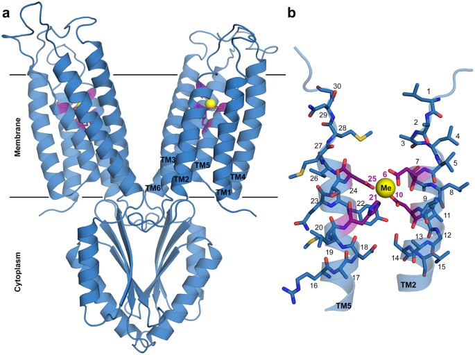
Transition metal binding selectivity in proteins and its correlation with  the phylogenomic classification of the cation diffusion facilitator protein  family | Scientific Reports
Transition metal binding selectivity in proteins and its correlation with the phylogenomic classification of the cation diffusion facilitator protein family | Scientific Reports
Encyclopedia | Free Full-Text | Metal Binding Proteins

RCSB PDB - 3EPV: X-ray Structure of the Metal-sensor CnrX in both the Apo-  and Copper-bound Forms
RCSB PDB - 3EPV: X-ray Structure of the Metal-sensor CnrX in both the Apo- and Copper-bound Forms

RCSB PDB - 1IMF: STRUCTURAL STUDIES OF METAL BINDING BY INOSITOL  MONOPHOSPHATASE: EVIDENCE FOR TWO-METAL ION CATALYSIS
RCSB PDB - 1IMF: STRUCTURAL STUDIES OF METAL BINDING BY INOSITOL MONOPHOSPHATASE: EVIDENCE FOR TWO-METAL ION CATALYSIS
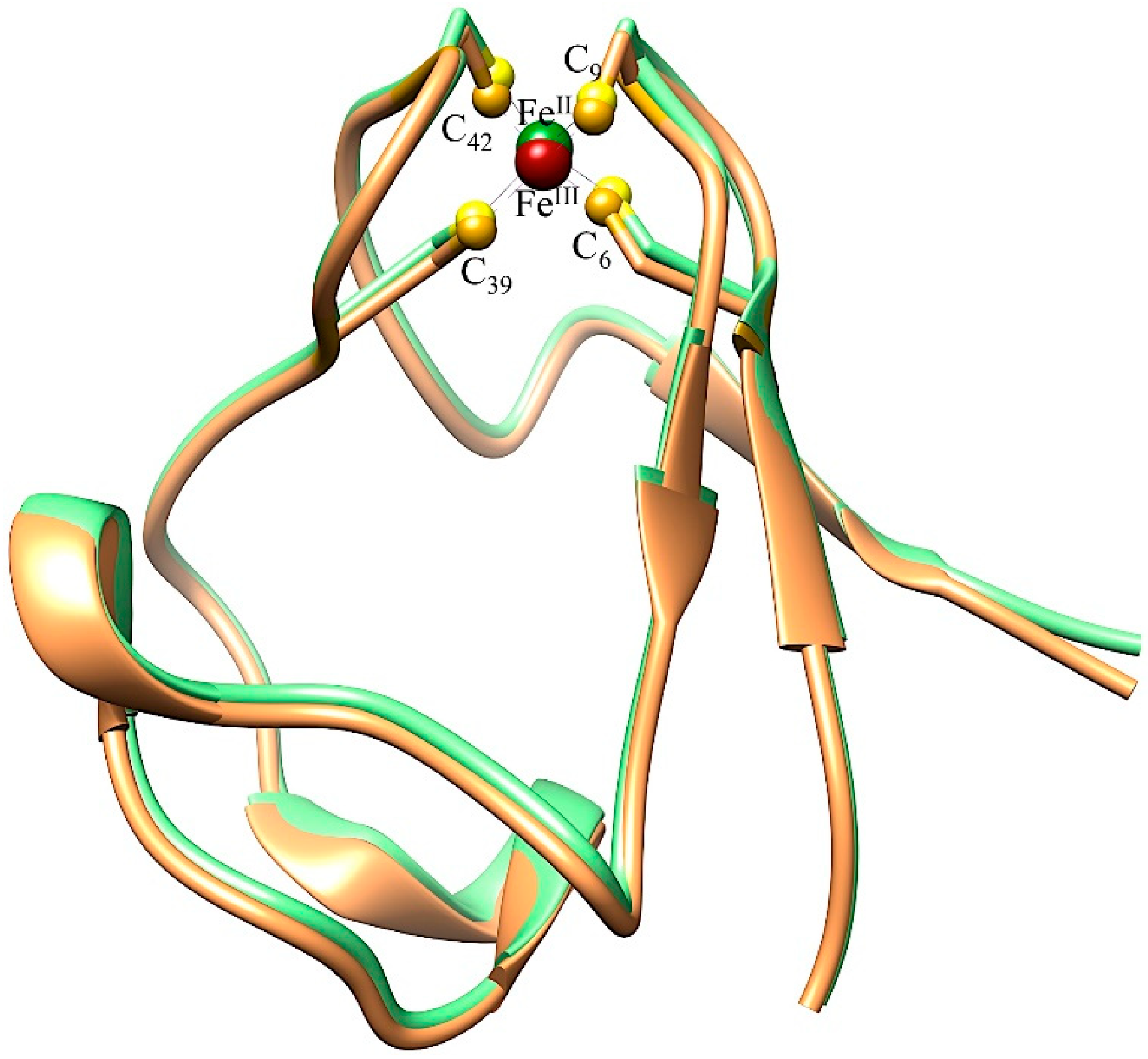
RCSB PDB - 1TUM: MUTT PYROPHOSPHOHYDROLASE-METAL-NUCLEOTIDE-METAL COMPLEX,  NMR, 16 STRUCTURES
RCSB PDB - 1TUM: MUTT PYROPHOSPHOHYDROLASE-METAL-NUCLEOTIDE-METAL COMPLEX, NMR, 16 STRUCTURES

BioChem | Free Full-Text | Native Protein Template Assisted Synthesis of  Non-Native Metal-Sulfur Clusters
BioChem | Free Full-Text | Native Protein Template Assisted Synthesis of Non-Native Metal-Sulfur Clusters
RCSB PDB - 2IUB: Crystal structure of a divalent metal ion transporter CorA  at 2.9 A resolution.
RCSB PDB - 2IUB: Crystal structure of a divalent metal ion transporter CorA at 2.9 A resolution.

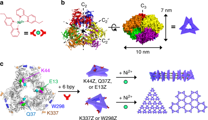
Diverse protein assembly driven by metal and chelating amino acids with  selectivity and tunability | Nature Communications
Diverse protein assembly driven by metal and chelating amino acids with selectivity and tunability | Nature Communications

InterMetalDB: A Database and Browser of Intermolecular Metal Binding Sites  in Macromolecules with Structural Information | Journal of Proteome Research
InterMetalDB: A Database and Browser of Intermolecular Metal Binding Sites in Macromolecules with Structural Information | Journal of Proteome Research

Welcome to MetalPDB
Welcome to MetalPDB

Welcome to MetalPDB
Welcome to MetalPDB

RCSB PDB - 1LU0: Atomic Resolution Structure of Squash Trypsin Inhibitor:  Unexpected Metal Coordination
RCSB PDB - 1LU0: Atomic Resolution Structure of Squash Trypsin Inhibitor: Unexpected Metal Coordination
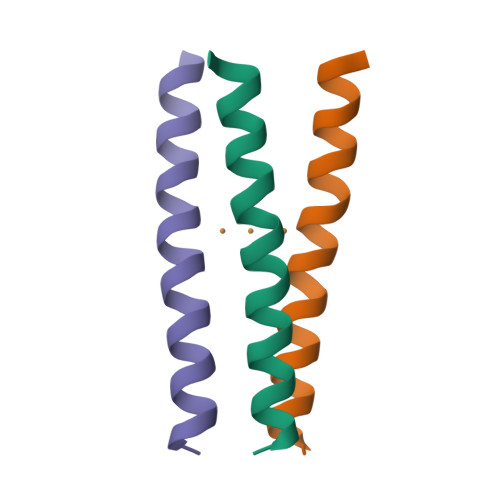
RCSB PDB - 1UT8: Divalent metal ions (zinc) bound to T5 5'-exonuclease

RCSB PDB - 3KSO: Structure and Mechanism of the Heavy Metal Transporter CusA

RCSB PDB - 6I1J: Selective formation of trinuclear transition metal centers  in a trimeric helical peptide
RCSB PDB - 6I1J: Selective formation of trinuclear transition metal centers in a trimeric helical peptide

RCSB PDB - 1XM5: Crystal structure of metal-dependent hydrolase ybeY from  E. coli, Pfam UPF0054
RCSB PDB - 1XM5: Crystal structure of metal-dependent hydrolase ybeY from E. coli, Pfam UPF0054

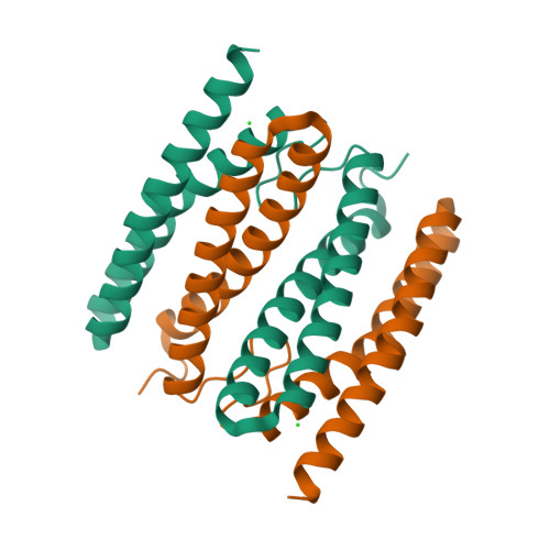
RCSB PDB - 7O89: sulerythrin without metals (apo-state)

Tools: Tools @ MetalWeb

RCSB PDB - 1MBX: CRYSTAL STRUCTURE ANALYSIS OF ClpSN WITH TRANSITION METAL  ION BOUND
RCSB PDB - 1MBX: CRYSTAL STRUCTURE ANALYSIS OF ClpSN WITH TRANSITION METAL ION BOUND

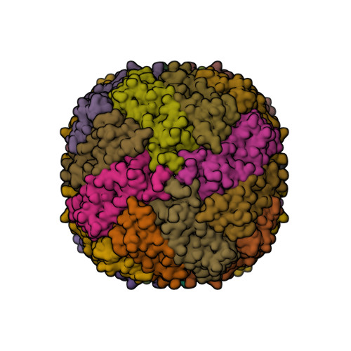
RCSB PDB - 5OUZ: Metal free structure of Y40F SynFtn

Plafoniera 4 luci - 264.340 Manta - Metal Lux | Lucilla Place

InterMetalDB: A Database and Browser of Intermolecular Metal Binding Sites  in Macromolecules with Structural Information | Journal of Proteome Research
InterMetalDB: A Database and Browser of Intermolecular Metal Binding Sites in Macromolecules with Structural Information | Journal of Proteome Research

RCSB PDB - 1AD7: NMR STRUCTURE OF METAL-FREE CONANTOKIN G, 1 STRUCTURE

RCSB PDB - 3KSO: Structure and Mechanism of the Heavy Metal Transporter CusA

Pymol illustration of Hg II S [Zn II N (H 2 O)](CSL9CL23H) 3 + (PDB... |  Download Scientific Diagram
Pymol illustration of Hg II S [Zn II N (H 2 O)](CSL9CL23H) 3 + (PDB... | Download Scientific Diagram