Home

Strumento Collana Riconoscimento gram stain smear Scaduto Rosso soddisfazione

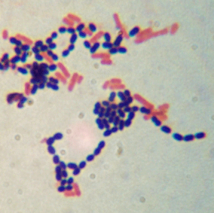
Dead leukocytes (Pus cells) in Gram Stained Smear

Welcome to Microbugz - Gram Stain

File:Gram Negative Rods in Gram Stained Smear of Culture.jpg - Wikimedia  Commons
File:Gram Negative Rods in Gram Stained Smear of Culture.jpg - Wikimedia Commons

Gram Stain (Cervical Smear) | NAOKON DIAGNOSTICS

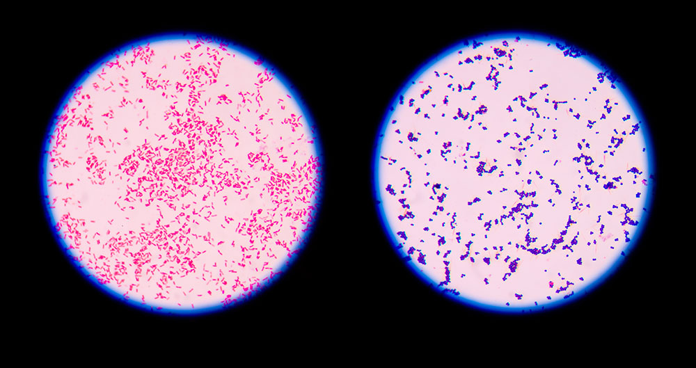
Gram Positive vs Gram Negative | Technology Networks

Wet Smear Gram Stain Undermicroscropy Showing Stock Photo 671716339 |  Shutterstock
Wet Smear Gram Stain Undermicroscropy Showing Stock Photo 671716339 | Shutterstock

READING OF A GRAM STAIN IN DIAGNOSTIC MICROBIOLOGY Dr.T.V.Rao MD

Gram Stain: Reference Range, Interpretation, Collection and Panels

Module 5.2: Microbial Stains – Clinical Veterinary Diagnostic Laboratory

VitroView™ Gram Stain Kit for Slide Smear (250 ml per bottle)

File:Bacteria and pus cells in a Gram stained smear of sputum.jpg -  Wikimedia Commons
File:Bacteria and pus cells in a Gram stained smear of sputum.jpg - Wikimedia Commons

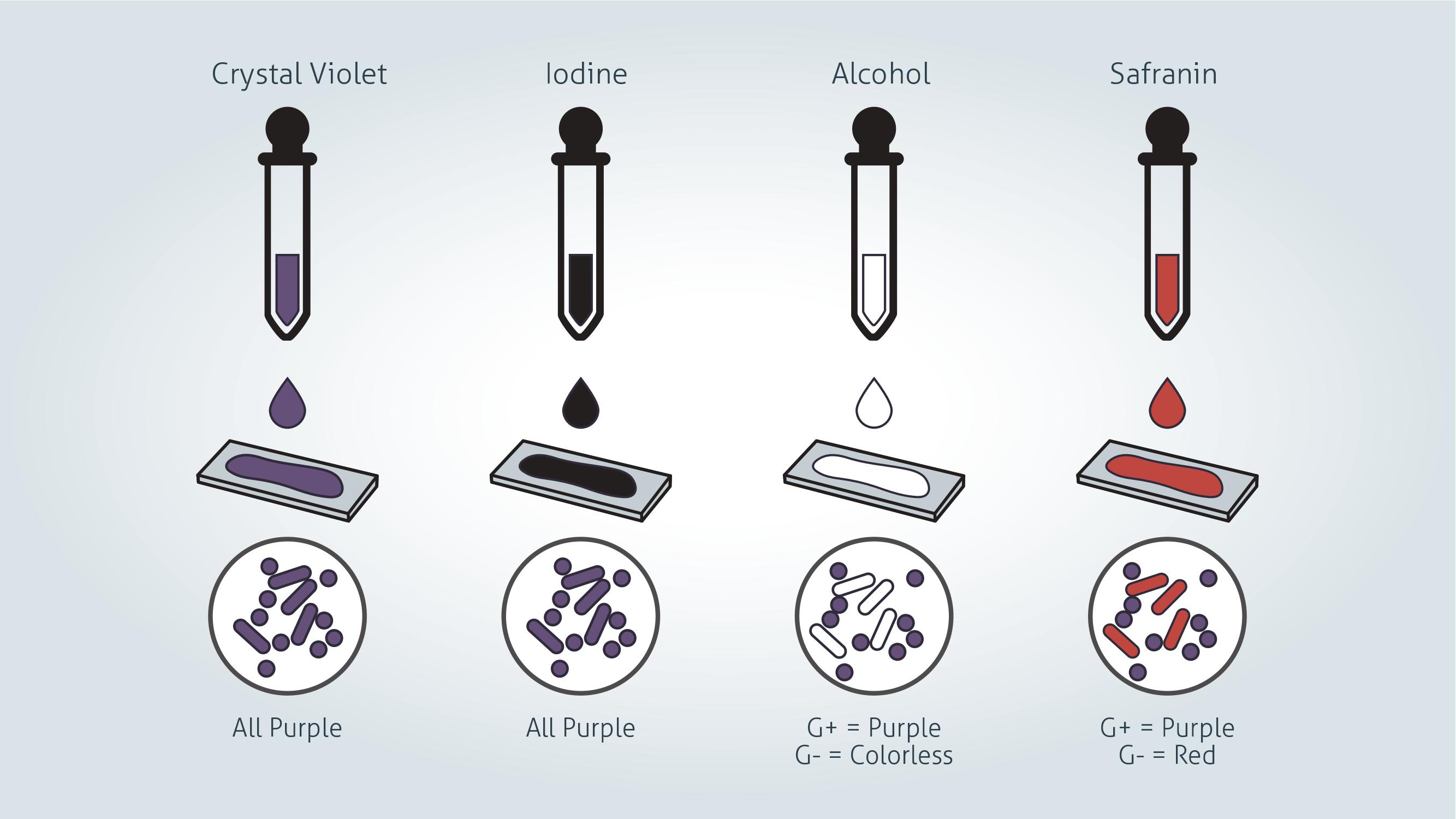
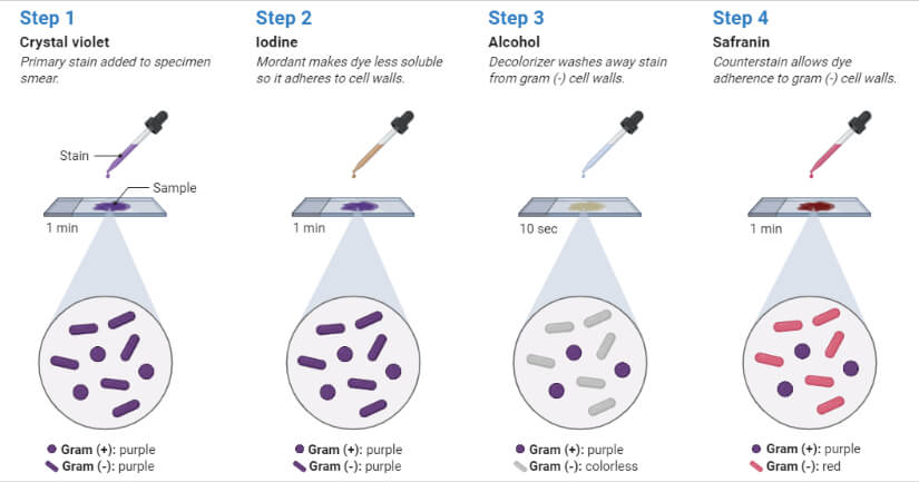
Gram Stain Protocols | ASM.org

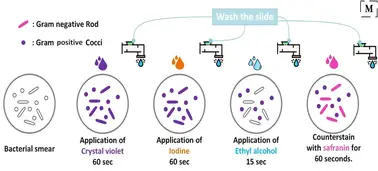
Gram staining - Procedure, mechanism, explanation

Gram-stained blood smear. Gram stain, also called the Gram method, is a...  | Download Scientific Diagram
Gram-stained blood smear. Gram stain, also called the Gram method, is a... | Download Scientific Diagram
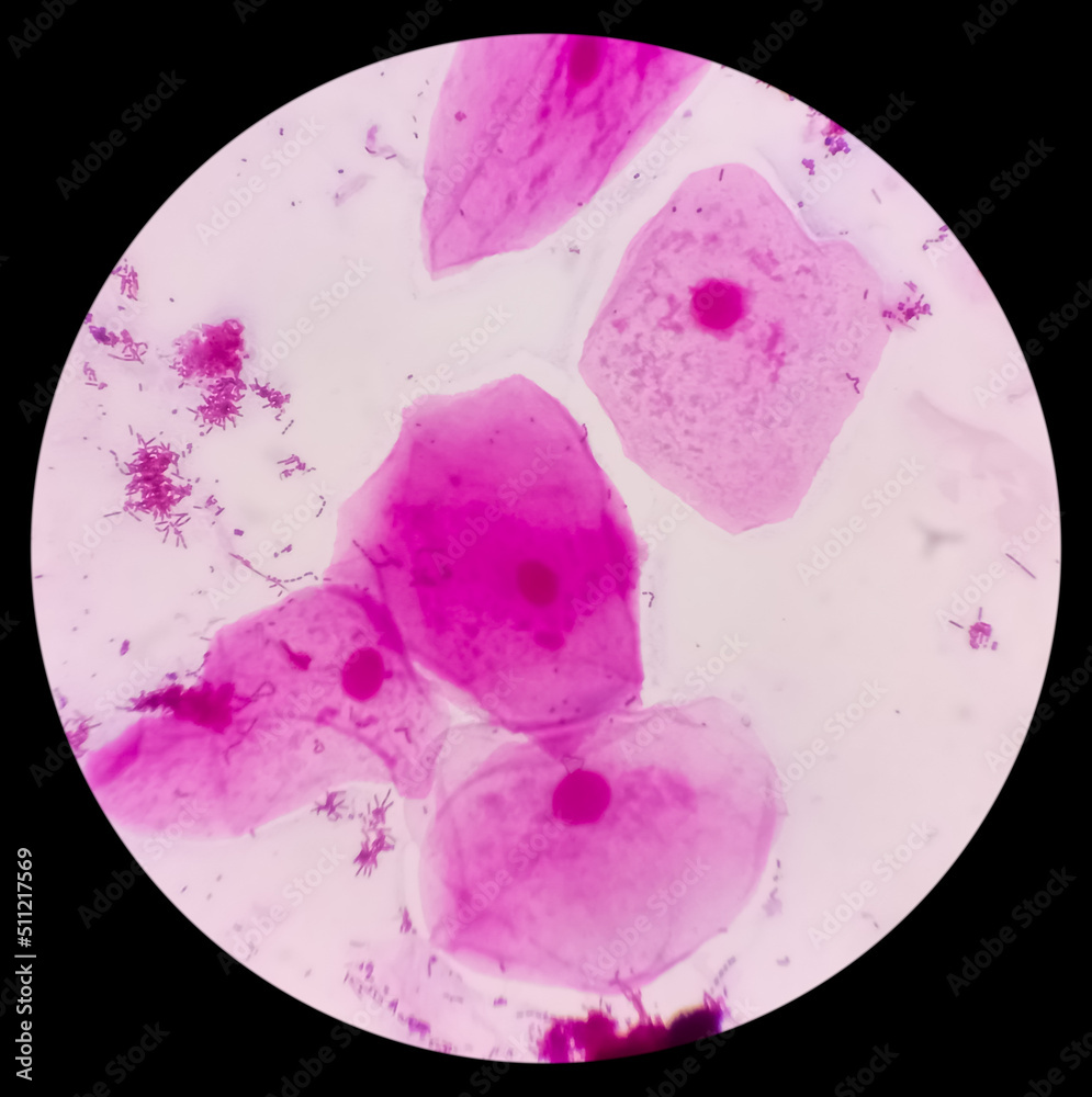
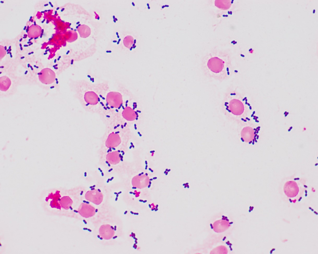
Foto Stock Prostatic Smear (PS) gram stain microscopic 40x show epithelial  cells. Large number of gram positive Diplococci and few gram negative rods  shape bacteria. | Adobe Stock
Foto Stock Prostatic Smear (PS) gram stain microscopic 40x show epithelial cells. Large number of gram positive Diplococci and few gram negative rods shape bacteria. | Adobe Stock

Gram Stain: Looking Beyond Bacteria to Find Fungi in Gram Stained Smear: A  laboratory guide for medical microbiology
Gram Stain: Looking Beyond Bacteria to Find Fungi in Gram Stained Smear: A laboratory guide for medical microbiology

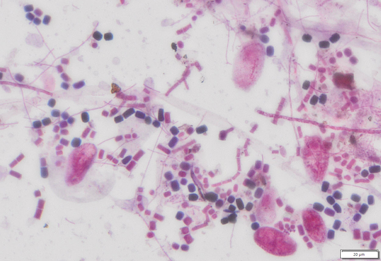
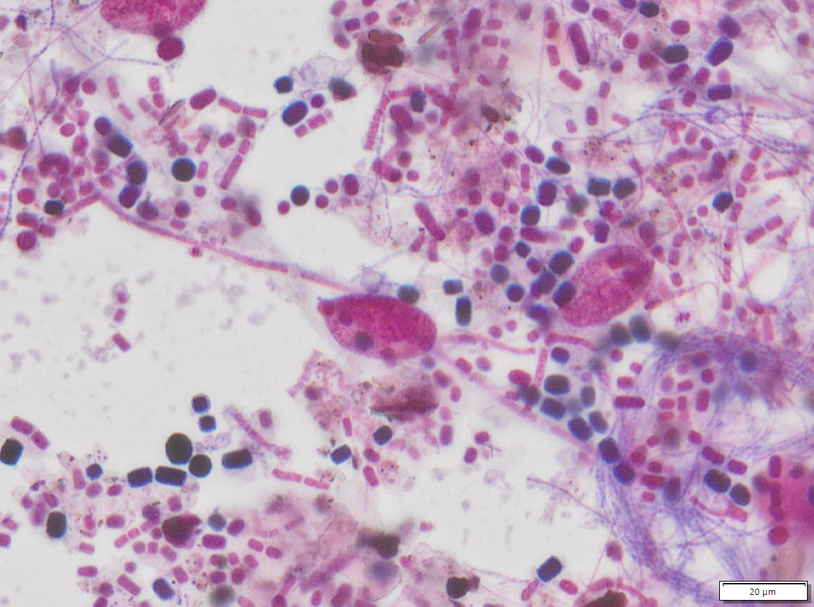
Large gram-positive organism. Gram stained smear | Download Scientific  Diagram
Large gram-positive organism. Gram stained smear | Download Scientific Diagram

VitroView™ Gram Stain Kit for Slide Smear (30 ml per Dropper bottle)

Gram Staining : Principle, Procedure and Results

Gram Staining- Principle, Reagents, Procedure, Steps, Results

Prostatic Smear Ps Gram Stain Microscopic Stock Photo 2053943189 |  Shutterstock
Prostatic Smear Ps Gram Stain Microscopic Stock Photo 2053943189 | Shutterstock

Gram-stained smear of pus sample. a The budding of the eosinophilic... |  Download Scientific Diagram
Gram-stained smear of pus sample. a The budding of the eosinophilic... | Download Scientific Diagram

Gram stain for microbiology student | PDF

How to Prepare the Perfect Gram Stain 2023